 Nano Aerosol Chamber In-Vitro Toxicity
Effects of inhaled nanoparticles to normal and diseased airway epithelia by in vitro technology
Chamber Characterization
NACIVT performance
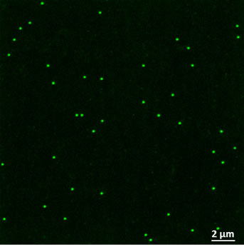
Distribution of polystyrene latex (PSL, yellow-green, 200 nm diameter) particles on Transwell® inserts after 0.5-h exposure in NACIVT: Left/Upper: Laser scanning micrograph shows the uniform distribution of deposited PSL particles. Right/Lower: The graph showing deposited particles as a function of the distance from the center of the Transwell® membrane demonstrates even distribution of PSL particles over the insert membrane except for the outermost zone, where considerably fewer particles were deposited. Read more (Jeannet et al., Nanotoxicology 9(1):34-42 (2015), DOI: 10.3109/17435390.2014.886739).
![]()
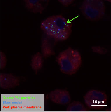
Deposited particles reach the cells: Laser scanning micrograph showing uptake of green fluorescent PSL particles (yellow-green, 200 nm diameter, arrow) by macrophages after 1-h exposure in NACIVT (Jeannet et al., Nanotoxicology 9(1):34-42 (2015), DOI: 10.3109/17435390.2014.886739).
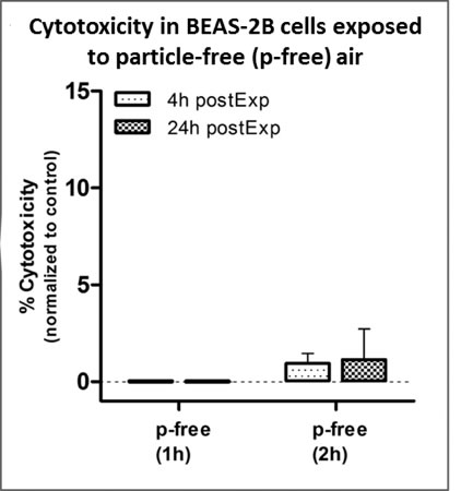
NACIVT is suitable for use with cell cultures. For example, exposure of BEAS-2B cells to particle-free air does not increase necrotic cell death (% cytotoxicity) (Jeannet et al., Nanotoxicology 9(1):34-42 (2015), DOI: 10.3109/17435390.2014.886739).
Adverse effects of NP aerosol exposure to normal and diseased epithelia
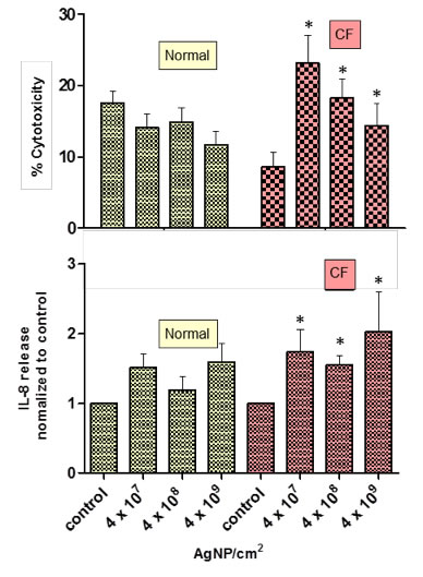
Cultures of fully differentiated normal and cystic fibrosis (CF) epithelia were exposed to three different concentrations of silver nanoparticles (AgNP) during maximally 1 hour. Cell analysis at 24 h after aerosol exposure revealed that CF epithelia responded more sensitively and at all doses to NP exposure than normal epithelia. For example, necrotic cell death (% cytotoxicity) as well as interleukin-8 (IL-8) release were significantly (*p < 0.05) increased compared to control values in CF but not in normal epithelia (Jeannet et al., Nanotoxicology, 2015, DOI 10.3109/17435390.2015.1049233).
Cell Analyses
After particle exposure, the biological effects induced by the particles in the cell culture can be analyzed for any biological marker of choice. We routinely assess the following parameters:
Endpoints
- Cytotoxicity
- LDH release
- Caspase-3 activity
- (Pro-)Inflammatory responses
- Cytokine release (IL-6, IL-8, MCP-1)
- Gene expression
(mRNA of IL-6, IL-8, MCP-1, HMOX-1, COX-2)
- Function of airway epithelia
- Barrier (Trans Epithelial Electric Resistance, TEER,
maintenance of air-liquid interface (ALI) - Defense (Ciliary beating, mucus production)
- Barrier (Trans Epithelial Electric Resistance, TEER,
- Morphology including cellular ultrastructure
- Light and electron microscopy
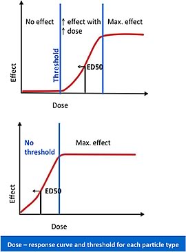
Hazard identification & characterization:
Dose - response relationship